The
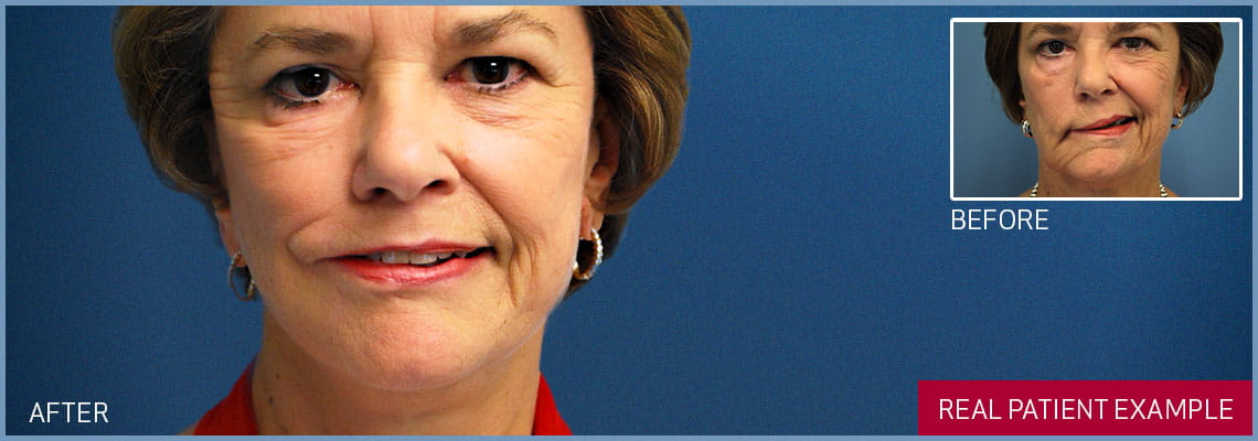
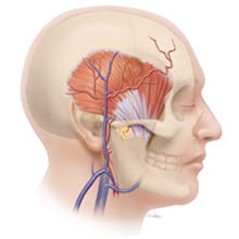
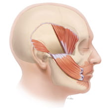
temporalis muscle is often chosen because its vector or angle of pull produces an upwardly oriented smile. The masseter muscle transfer creates a more horizontal movement at the corner of the mouth. Utilizing the temporalis and masseter transfers in combination has the potential to produce a multidirectional smile.
Muscle transfer procedures are often reserved for individuals whose age, general medical condition or previous surgeries prohibit more complex microsurgical procedures. These procedures can also be utilized to augment facial motion in individuals who have made a partial recovery after facial nerve paralysis.
There are three principle ways of transferring the temporalis muscle:
- Segmental muscle turn-over
- Orthodromic muscle flap with fascia lata graft.
- Lengthening myoplasty
The exact technique selected is guided by a complex series of variables including the degree of facial weakness.
Technique
Segmental muscle turn-over
The temporalis muscle transfer is performed through a face lift style incision that is created in front of the ear and extends into the temple. The central segment of the temporalis muscle is released on three sides leaving its base attached.
The dense leathery tissue encasing the muscle (fascia) is unfurled to extend its reach. The temporalis muscle segment is then tunneled beneath the skin of the cheek and secured to the upper lip. The temporal void created by borrowing the muscle segment can be filled by rearranging local tissues or utilizing a number of biological or synthetic implants.
Orthodromic Temporalis Muscle Flap with Fascia Lata Graft
An incision is created in the hair bearing region of the temple and extended for a limited distance in front of the ear. The tendinous portion of the temporalis muscle is now freed from its attachment to the jaw. A graft of leathery tissue (fascia lata) obtained from the thigh is utilized to bridge the free end of the muscle to the lips and corner of the mouth. When the teeth are clenched the fascial graft transfers the force of the temporalis muscle to the corner of the mouth creating a voluntary smile. With diligent practice motivated individuals can develop a smile that is reflexive and biting is no longer required.
Lengthening Temporalis Myoplasty
A scalp incision is created in a zig-zag fashion similar to a cosmetic brow lift. The temporalis muscle is mobilized from its attachments to the underlying cranial bone and its tendious insertion onto the jaw is released. Intermittently, the cheek bone (zygomatic arch) may be temporarily released to facilitate the procedure and is restored to its native position at the completion of the surgery. A limited incision is now created in the natural fold that extends from the base of the nose to the corner of the mouth (nasolabial fold).
Through this aperture the mobilized temporalis flap can be sutured to the corner of the mouth. The proximal muscle is re-anchored in the scalp region. With clenching of the teeth the force of the muscle is re-directed to the corner of the mouth creating a voluntary smile. Again, with diligent practice many individuals through the process of neural plasticity with learn to create a smile that is independent of jaw clenching.
