
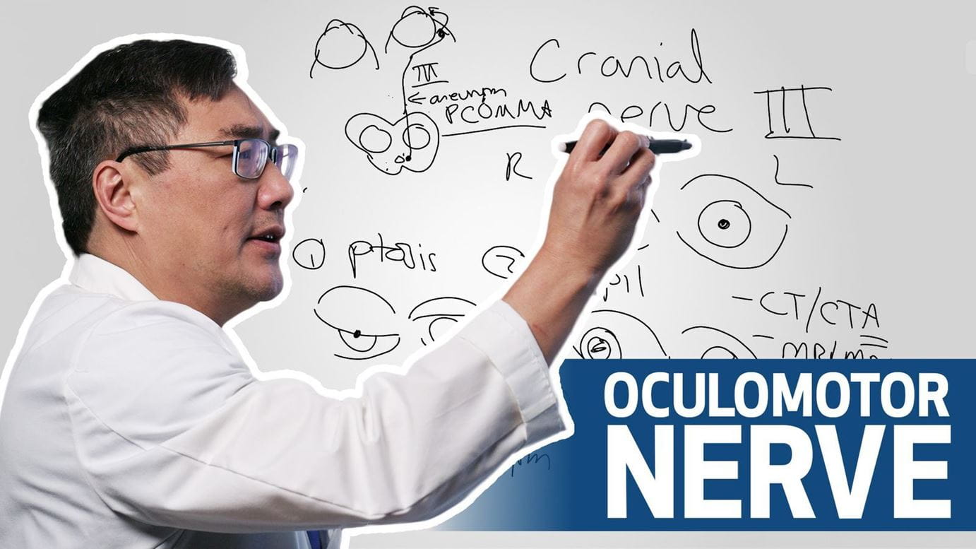

Cranial nerve III palsy can present through eyelid drooping, pupil changes and eye misalignment — urgent evaluation is critical.
Article Highlights
- Cranial nerve III palsy presents with ptosis, pupillary dilation and ocular misalignment, often producing a “down and out” eye.
- Any pupil-involved third nerve palsy should be presumed aneurysmal (typically posterior communicating artery) until proven otherwise.
- Untreated aneurysms linked to CN III palsy carry a high risk of rupture and mortality, making this an emergency evaluation.
- Initial workup prioritizes non-contrast CT followed by CT angiography, with MRI/MRA used to evaluate alternative etiologies if negative.
- Beyond aneurysm, causes include ischemic, compressive and structural lesions (e.g., brainstem infarct, cavernous sinus mass or orbital pathology), requiring comprehensive neuroimaging.
In Houston Methodist's latest whiteboard video, neuro-ophthalmologist Dr. Andy Lee breaks down the signs, causes and urgency of diagnosing cranial nerve III palsy — a condition that can signal life-threatening aneurysms if missed.
"The third cranial nerve supplies the eye movement, the pupil and the lid, so we have to look at all three when we're looking at diagnosing third nerve palsy," says Dr. Lee, chair of the Blanton Eye Institute at Houston Methodist Hospital.
Patients typically present with three hallmark signs: drooping eyelid (ptosis), a dilated pupil and eye misalignment. Because the nerve controls several muscles that move the eye, paralysis can force the eye downward and outward.
"Ptosis plus misalignment plus pupil are the three components of third nerve palsy," explains Dr. Lee in the video.
The condition is particularly concerning because of its link to aneurysms of the posterior communicating artery.
“It's really important that you know what a cranial nerve III palsy looks like. It's an emergency evaluation you can't afford to treat like a normal one.”
Dr. Andy Lee
"A pupil-involved third nerve palsy is an aneurysm … unless or until proven otherwise," Dr. Lee stresses. "If we treat it, it's a cure. If we let it rupture, half those people die at the scene. That's why the workup is always treated as an emergency."
Houston Methodist experts recommend beginning with a non-contrast CT scan (PDF) to rule out hemorrhage, followed immediately by CT angiography to look for aneurysm. If those are negative, MRI and MR angiography are performed to identify other potential causes such as brain stem infarct, cavernous sinus mass or orbital lesion.
By educating physicians on the classic features and critical imaging pathway, Dr. Lee says he hopes to highlight both the diagnostic challenge and the opportunity to save lives.