Masdeu Lab

11907

Citations

45

h-index

About Our Lab

Using the tools of human neuroimaging, including magnetic resonance imaging (MRI), positron emission tomography (PET) and electroencephalography (EEG), the neuroimaging lab aims to clarify the neurobiology and improve the treatment of the following disorders causing cognitive impairment.
 
 
Alzheimer’s disease
  • Timing of beta-amyloid and tau deposition in preclinical stages
  • Genetic effects
  • Effect of treatment
  • Correlation of tau imaging with postmortem tau quantification

Frontotemporal dementia
  • Genetic underpinning of regional involvement patterns
  • Neuroimaging correlates of social cognition

Traumatic brain injury
  • Amyloid and tau imaging in acute and chronic concussion
  • Timing and brain localization of tau deposition in patients with repeated head trauma

Schizophrenia
  • Anti-synaptic antibodies and their effect on functional connectivity

Working with the MRI and PET facilities in the Translational Imaging Core of the Houston Methodist Research Institute and with the collaboration of a number of external researchers, the lab has utilized and implemented the following techniques:

MRI-related
  • Structural MRI
  • BOLD MRI
  • Activation
  • Resting, with functional connectivity
  • Diffusion tensor imaging (DTI)
  • Susceptibility weighted imaging (SWI)
  • Arterial spin labeling (ASL) 

PET
PET Radiopharmaceuticals
  • [18F] Fludeoxyglucose
  • [11C] Pittsburgh compound B
  • [18F] T807 (AV-1451)
  • [18F] Fluoro-L-DOPA
  • [11C] PBR28


Scientific Discoveries 


There is an inverse regional relationship between the amount of tau and metabolism in the brain
Our laboratory was the first to report that areas of the brain that have abnormal hyperphosphorylated tau deposition have decreased metabolism, with a high anatomical concordance. In the healthy brain, the protein tau stabilizes neurotubules and is therefore essential for normal neural function. However, in Alzheimer’s disease and other neurodegenerative disorders, tau becomes abnormally hyperphosphorylated, dysfunctional and misfolded, constituting the neurofibrillary tangles observed neuropathologically in Alzheimer’s disease and other tauopathies [1]. We discovered that there is an inverse regional relationship between the amount of tau in the brain and the metabolism in the same region: regions with high tau have low metabolism and vice-versa [2]. By contrast, this relationship does not apply to amyloid deposition: areas with high amyloid load (for instance, the calcarine cortex in Fig 1) may have normal metabolism. An image from the paper reporting this finding was used to illustrate the cover of the article where this research was published [2]. This finding has critical implications: because metabolism indicates the activity of the various regions of the brain, and it is depressed precisely in those areas with a high load of tau, but not amyloid, this finding suggests that it is tau that is critical, rather than amyloid, to the development of clinical impairment in the disease [1]. It also suggests that monitoring the spreading of tau will be a powerful biomarker for the progression of the disease and that tau imaging can be used to monitor the effectiveness of potential new therapies, perhaps using patient samples much smaller than needed when cognitive impairment is used as an outcome, as currently done.


This finding does not mean that therapeutic approaches targeting amyloid are useless. They may well work in the pre-tau stage of the disease, when people predisposed to developing Alzheimer’s are still asymptomatic, but they are unlikely to help once tau has spread to neocortical networks. This assumption is supported by the recent announcements of the failure of amyloid-removing clinical trials at the early Alzheimer’s disease stages.


Fig 1. 18F-FDG, 18F-florbetapir, and 18F-AV-1451 PET superimposed to native MRI. Yin-yang relationship between tau and metabolism in the brain of a patient with Alzheimer’s disease. Multimodality positron emission tomography (PET) images are superimposed to native MRI. Primary sensory-motor areas (asterisks), as well as the primary visual (striatal cortex) and auditory (Heschl’s gyrus) regions (arrowheads) have normal metabolism and no tau deposition. Areas with high tau deposition (e.g. inferior parietal lobule, arrows) have decreased metabolism. This relationship does not apply to amyloid: notice high amyloid uptake in the calcarine cortex on the 18F-florbetapir scan (central column, arrowheads). AVID Radiopharmaceu-ticals provided the precursor for 18F-AV-1451.


Tau propagates within  natural brain networks (“neurons that wire together degenerate together”)
Tau propagates within  natural brain networks (“neurons that wire together degenerate together”).

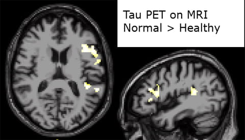
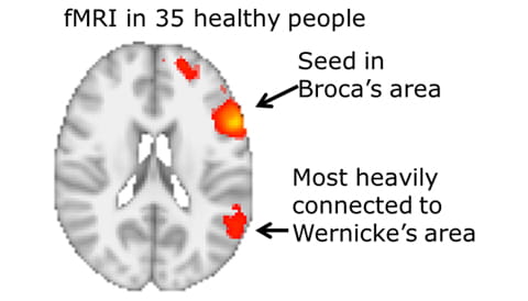
Our  laboratory has produced the first direct in  vivo demonstration that tau spreads across the language network. We  evaluated with tau (18F-AV-1451) PET a group of patients with the  nonfluent variant of primary progressive aphasia (nfvPPA), a progressive  neurodegenerative disorder that is, like Alzheimer’s disease, tau-dependent [3], and a  group of healthy controls. Both patients and controls were amyloid-negative,  and did not differ significantly in age and sex. PET images from 80 to 100 min  were averaged, and standardized uptake value ratios were calculated using the  mean activity in the cerebellar gray matter as the reference region.  Additionally we determined functional connectivity with BOLD MRI in a group of  healthy volunteers group-matched to the patients, using as seed the volume of  the largest tau cluster in the nfvPPA group.

On  voxel-wise analysis and compared to controls, patients with nfvPPA had  increased tau uptake (p < 0.001) in two major clusters. The larger cluster  was in Broca’s area. The smaller cluster was located in the planum temporale of  the superior temporal gyrus, extending to the supramarginal gyrus (Wernicke’s  area). As expected, MRI functional connectivity from the location of the larger  tau cluster was greatest with a region located in the same area as the smaller  tau cluster [4]. 


Fig  2. Tau, detected with 18F-AV-1451 PET in patients with non-fluent  primary progressive aphasia, propagates in the language network. Significant  areas of increased 18F-AV-1451 deposition in patients compared to  age and sex-matched healthy controls (p < 0.001). The largest cluster is in  the premotor region of the frontal lobe (Broca’s area). The smaller cluster is  in the planum  temporale (Wernicke’s  area), immediately posterior to the transverse temporal gyrus of Heschl.


Fig  3. Functional MRI connectivity map of the language network. Using  resting functional MRI data from healthy volunteers matched to the patients  whose data is shown in Fig 2, a seed was placed in the region with the greatest  tau deposition in the patients. This region was shown to connect most strongly  with precisely the region of the brain containing the second strongest tau peak  in patients, in Wernicke’s area.


Regions of “off-target-binding” in 18F-AV-1451 PET of healthy older people have increased permeability in older as compared to younger persons
In older healthy subjects, [18F]AV-1451, which is supposed to bind to hyperphosphorylated tau, also seems to bind to regions (putamen and other nuclei) known not to have abnormal tau deposition on neuropathology (Fig 4). This so-called “off-target” binding has caused reports of abnormal tau deposition in tau PET studies of people who were actually normal. Our aim was to determine whether apparently increased specific [18F]AV-1451 binding in areas unlikely to harbor hyperphosphorylated tau, such as the substantia nigra, globus pallidus and putamen, of older subjects could be related to greater vascular permeability of these regions in older subjects as compared to younger ones.

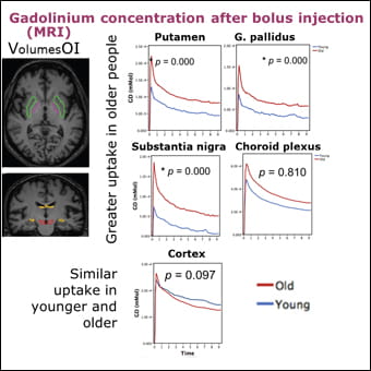
For this purpose we measured dynamic [18F]AV-1451 uptake over a three-hour period in younger (23±2.1 years of age) and older (68.8±7.6 years) healthy subjects. In the same subjects we obtained dynamic gadolinium concentrations before and after a bolus injection of gadolinium, a large molecule commonly used to assess the permeability of the blood-brain barrier, by performing a pre-contrast 3D T1 map (five flip angle acquisitions) followed by a dynamic contrast enhanced MRI (DCE-MRI). During and after infusion, we acquired 180 consecutive T1-weighted volumes (3.4 sec per volume) over 10 minutes. Non-displaceable binding potential (BPND) of [18F]AV-1451 and permeability parameters (Ktrans) of gadolinium were analyzed by volume-of-interest methods and compared using Spearman’s correlation coefficients.

We found that there was greater BPND of [18F]AV-1451 in the globus pallidus and putamen (but not in cerebellum or cerebral cortex) of older as compared to younger subjects (Fig 4). Gadolinium concentration and permeability (Ktrans) was similar in the cerebellum, temporal cortex, and choroid plexus of younger and older subjects, but there was higher permeability in the substantia nigra, globus pallidus, and putamen (all p<0.05) of older as compared to younger subjects (Fig 5). Correlations between BPND and Ktrans were significant for substantia nigra, globus pallidus and putamen (all p<0.05) [5].


Fig 4. Tau ([18F]AV-1451) PET shows greater uptake in “off-target” regions in older than in younger people. [18F]AV-1451 PET superimposed to native MRI in a younger (left) and an older (right) adult. Uptake in these regions has been misinterpreted in some publications to represent tau deposition, when several neuropathological studies have failed to show abnormal tau in these regions. 


Fig 5. Gadolinium concentration was greater in “off-target” regions in older than in younger people. Anatomic panels on the left show the position of the volumes of interest for the graphs depicted on the right. Each graph shows gadolinium concentration in the Y axis and time post bolus injection in the X axis.


In summary, our work showed that increased vascular permeability in substantia nigra, pallidum and putamen of healthy older subjects could underlie some of the regional differences in [18F]AV-1451 uptake observed in older as compared to younger healthy subjects. These data are most relevant for the correct interpretation of the findings present in [18F]AV-1451 PET scans. This compound is now widely used to measure abnormal tau at all stages of the Alzheimer’s disease neurobiological continuum.


Most Recent Publications on Alzheimer’ Disease and Related Disorders (cited above):

  • Masdeu, J. (2016) Misfolded tau, Alzheimer's bogeyman, exposed. JAMA Neurol in press.
  • Pascual, B. and Masdeu, J.C. (2016) Tau, amyloid, and hypometabolism in the logopenic variant of primary progressive aphasia. Neurology 86, 487-488.
  • Masdeu, J.C. and Pascual, B. (2016) Genetic and degenerative disorders primarily causing dementia. Handb Clin Neurol 135, 525-564.
  • Pascual, B., Zanotti-Fregonara, P., Funk, Q., Rockers, E., Pal, N., Yu, M., . . . Masdeu, J. (2017) Nonfluent variant of primary progressive aphasia: Tau deposition in the language network. In Human Amyloid Imaging Conference.
  • Pascual, B., Rockers, E., Bajaj, S., Yu, M., Karmonik, C., Xue, Z., and Masdeu, J. (2016) Older healthy people have increased vascular permeability in regions showing “off-target” [18F]AV-1451 uptake. In Alzheimer's Association International Conference.
Research News
doctor holding a tablet with a chest scan on it's screen
Personalized Treatment Strategies for Lung Cancer
Houston Methodist researchers identified a novel and innovative marker to guide Immune Checkpoint Inhibitor Therapy for Lung Cancer.
woman sitting on an examination table in a doctor's office
Trailblazing New Perspectives: Treating Gynecological Cancers
The goal of this new study was to evaluate the feasibility of a phase three, multi-center randomized clinical trial comparing the efficacy of minimally invasive surgery with that of the traditional open method approach (laparotomy) in treating epithelial ovarian cancer.
arm in blood pressure testing machine
Getting to the Heart of Hypertension Disparities
Researchers address social risk factors for high blood pressure through a national study.
Evolving Treatment Beyond Pharmaceuticals
First-of-its-Kind Study Explores Novel Prescription Digital Therapeutic
An Unusual Presentation of Glioblastoma
Andrew Lee, MD, Herb and Jean Lyman Centennial Chair in Ophthalmology, recently published one of the first-ever reported cases of progressive vision loss as the presenting manifestation of recurrent GBM with secondary LMS to the optic chiasm.
Engaging T cell Exhaustion for Solid Tumor Immunotherapies
Houston Methodist researchers investigated T cell exhaustion in murine models of prostate cancer and melanoma to explore possible advancements in immunotherapies for solid tumors.
Fighting a Deadly Duo
Investigating therapeutics to fight deadly TB/HIV coinfections.
Houston Methodist, Rice University launch groundbreaking Digital Health Institute
This initiative builds on ongoing collaborations between Houston Methodist and Rice University to transform the future of health care.
Stage I Nodal Marginal Zone Lymphoma Treatment Landscape
Houston Methodist researchers compared systemic therapy and radiation therapy in Stage I Nodal Marginal Zone Lymphoma Patients
2024 Digital Health Workshop Seed Fund Award Winners Announced
Houston Methodist researchers and their collaborators from Rice University have been awarded grants from the 2024 PATHS-UP Digital Health Workshop.
Pilot Study Offers Insight Into Alzheimer’s Research Participants Perceptions and Self-Efficacy
In a Houston Methodist study, Alzheimer’s disease research participants showed a high interest in genetic testing and provided valuable insights that can improve how test results are disclosed.
Breast Cancer Cells
Nanomedicine Makes Big Strides in the Fight Against Breast Cancer
Researchers deliver immunotherapy directly into triple-negative breast cancer tumors with nanofluidic implants, achieving tunable and sustained dosing of immunotherapeutics with high anti-tumor activity.
headshot of Constance Mobley smiling
Liver Failure Avengers Houston Methodist Researchers First in World
Under the leadership of Constance Mobley, MD, PhD, FACS, researchers have successfully performed a first-in-human miniature liver hepatocyte transplant to change a patient’s lymph nodes into ectopic miniature livers.
Predicting the Future of Cancer Treatment

Houston Methodist faculty developed a multiscale mechanistic model to further investigate the role of miR-155 in non-small cell lung cancer and predict clinical efficacy based on preclinical data.

Taking a Closer Look at West Nile Neuroimaging

A case report reviewing West Nile neuroinvasive disease imaging characteristics and the differential diagnosis of acute leukoencephalopathy highlights the usefulness of neuroimaging in WNND diagnosis in the absence of CSF markers.

Ending Organ Transplant Rejection

Studies at Houston Methodist suggest a novel strategy that may potentially eliminate or diminish transplant rejection, autoimmune diseases, and the need for immunosuppressive drugs

Location, Location, Location is Key to Pancreatic Cancer Prognosis

Molecular alterations found in pancreatic ductal adenocarcinoma samples may have potential therapeutic implications

Donate to Houston Methodist

With your support, Houston Methodist provides exceptional research, education, and care that is truly leading medicine.

Donate Now